Cellular & Preclinical Models
Overview
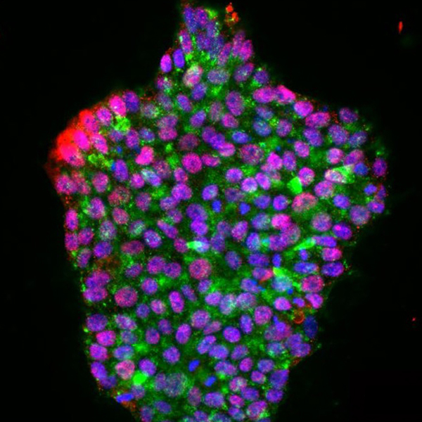
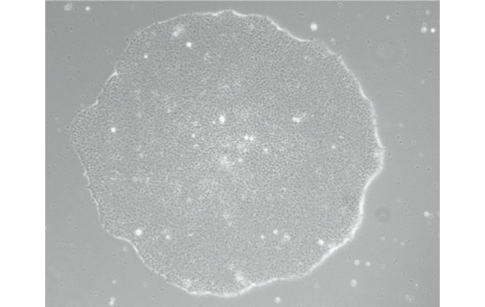
In vitro models are essential for cancer research, providing controlled systems to study the molecular mechanisms behind cancer development, progression, and response to therapy. Immortalized cell lines, capable of growing indefinitely, and organoids derived from tumor tissue help researchers investigate how cancer arises, test new treatments, and explore drug resistance.
The Cellular and Preclinical Models Facility provides the infrastructure and expertise needed to develop and maintain these models under strictly controlled conditions. Ensuring reproducibility is crucial, allowing researchers to generate reliable data, while rigorous safety measures are in place, particularly when using viruses to modify the genetic makeup of cells.
By supporting the creation and use of advanced cellular and preclinical models, the facility plays a fundamental role in advancing cancer research and improving future therapies.
The PCM Core Facility offers tailored introductory courses for young students, providing both theoretical and hands-on training. These courses are essential in equipping researchers with the necessary skills to effectively utilize the facility’s resources and methodologies.
In terms of infrastructure, the Cellular and Preclinical Models Facility is equipped with multiple dedicated spaces, including seven BSL-1 rooms and four BSL-2 rooms, ensuring the safe handling of cells in compliance with biosafety standards.
To ensure proper use of these spaces, new scientists must complete an introductory course, pass an assessment, and formally accept all relevant procedures before accessing the cell culture rooms.
Core Services Provided
- Management of the Institutional Cell Bank: The facility oversees a collection of over 300 cell lines, primarily cancerous, from human and animal origins. These lines are sourced from major international cell banks (ATCC, DSMZ, NCI, ECACC) and are available to all IFOM scientists.
- Cell Line Characterization & Quality Control:Each cell line undergoes rigorous quality checks, including Mycoplasma detection (enzymatic test and PCR), authentication by STR profile analysis, growth curve assessment, and basal resistance testing (puromycin, hygromycin, G418).
- Optimization of Transfection & Electroporation Protocols:The facility continuously refines protocols for frequently used cell lines to enhance efficiency and reproducibility.
- Quality Control on Private Collections & Primary Material: Quality checks are also conducted on primary samples and cell lines from individual research groups
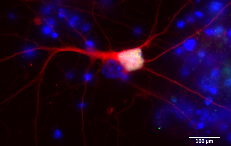
- Human iPSC Support:The facility assists research groups lacking expertise in stem cell handling by either conducting experiments on their behalf or providing training.
- Mass Culture & Clonal Selection: Services include large-scale hybridoma culture and clonal selection following cell line transfection or transduction.
- Media & Reagent Preparation & Distribution: The facility selects and tests sera to maintain batch consistency. Common cell culture reagents are pre-aliquoted and stocked in each room for easy access.
- Plasmid Preparation for Lentiviral Production: This includes preparation and distribution of plasmids as well as daily plating of HEK293T cells for lentiviral production.
- Regulatory Support: Assistance in completing MOMG employment notifications for the Ministry of Health.
Collaboration with Athena
Since 2021, the Facility has been supporting scientists in the:
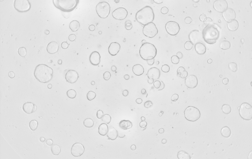
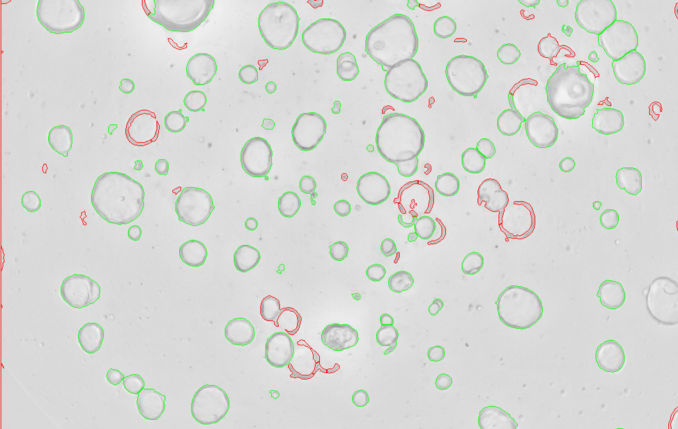
- Generation of CRC organoids.
- Production of conditioned media (including Noggin, R-spondin, and WNT) .
- Creation of Patient-derived organoids (PDOs) from human samples.
Each cell culture room is equipped with 8-10 hood places, 10-12 incubators and hosts roughly 20 researchers. For the handling of hazardous substances dedicated cytotoxic hoods are available. Quarantine areas (hoods and incubators) are available for newly arrived cell lines and freshly isolated primary material.
To further support a wide range of cell culture applications—from routine counting to live-cell imaging and specialized cell processing—the Cellular and Preclinical Models Core Facility is equipped with state-of-the-art instrumentation. This advanced setup ensures precise, reproducible conditions for cutting-edge research in cancer biology and beyond.
- Cell Counters (Beckman Multisizer 2, Thermo Countess I & II, Logos Bios LUNA)
Ensure accurate cell quantification and viability assessment for reproducible experimental results. - Electroporators (Thermo NEON, Nepagene NEPA21)
Facilitate the introduction of nucleic acids or proteins into cells through high-efficiency electroporation. - Tissue Dissociator (Miltenyi GentleMACS Octo)
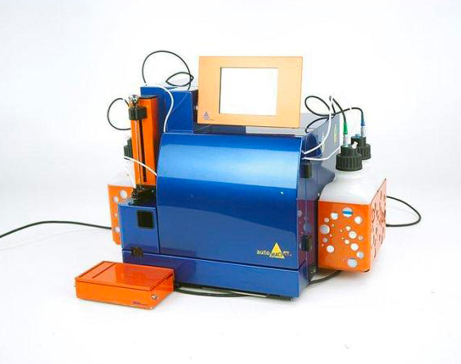
Enables the gentle and automated dissociation of tissues into single-cell suspensions, essential for downstream applications such as cell sorting and culture. - Magnetic Cell Separator (Miltenyi AutoMACS)
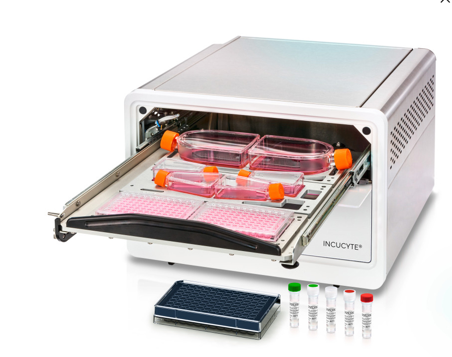
Allows for fast and efficient magnetic separation of specific cell populations for immunological and molecular studies. - Live-Cell Analysis System (Sartorius Incucyte S3 & S5)
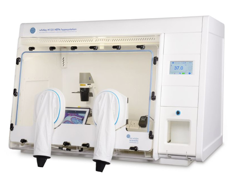
Provides real-time imaging and analysis of cell proliferation, morphology, and behavior under controlled conditions. - Hypoxia Stations (Don Whitley H45 & H135)
Maintain low-oxygen environments to simulate physiological or tumor microenvironment conditions, critical for hypoxia-related studies.
Facility Acknowledgment
According to our Core Facilities Guidelines, if Facility services were used in work described in a publication, the Facility must be acknowledged, citing both the Facillity name and its Research Resource Identifier (RRID). A typically acknowledgement might look like:
The research described was supported by the IFOM Cellular and Preclinical Models Core Facility (RRID: SCR_026864)
Co‑Authorship
If a staff member of an IFOM or Cogentech Core Facility contributes substantially to a published work, they should be included as a co‑author.
More information is available in Recommended guidelines and Acknowledging and citing core facilities
Examples of substantial contributions that may justify co‑authorship include:
- Contribution to experimental design or optimization
- Development or significant adaptation of methods or protocols
- Advanced data analysis and interpretation
- Intellectual input influencing the scientific direction of the study
Routine technical support or standard service provision generally warrants acknowledgment rather than co‑authorship.
-
PMID: 37855660
Leveraging Tissue-Specific Enhancer-Target Gene Regulatory Networks Identifies Enhancer Somatic Mutations That Functionally Impact Lung Cancer.
Hariprakash JM, Salviato E, La Mastra F, Sebestyén E, Tagliaferri I, Silva RS, Lucini F, Farina L, Cinquanta M, Rancati I, Riboni M, Minardi SP, Roz L, Gorini F, Lanzuolo C, Casola S, Ferrari F. Cancer Res. 2024 Jan 2;84(1):133-153. doi: 10.1158/0008-5472.CAN-23-1129. PMID: 37855660 Free PMC article. -
PMID: 38326317
N2FXm, a method for joint nuclear and cytoplasmic volume measurements, unravels the osmo-mechanical regulation of nuclear volume in mammalian cells.
Pennacchio FA, Poli A, Pramotton FM, Lavore S, Rancati I, Cinquanta M, Vorselen D, Prina E, Romano OM, Ferrari A, Piel M, Cosentino Lagomarsino M, Maiuri P. Nat Commun. 2024 Feb 7;15(1):1070. doi: 10.1038/s41467-024-45168-4. PMID: 38326317 Free PMC article. - PMID: 36293286
The PSI Domain of the MET Oncogene Encodes a Functional Disulfide Isomerase Essential for the Maturation of the Receptor Precursor.
Altintas DM, Gallo S, Basilico C, Cerqua M, Bocedi A, Vitacolonna A, Botti O, Casanova E, Rancati I, Milanese C, Notari S, Gambardella G, Ricci G, Mastroberardino PG, Boccaccio C, Crepaldi T, Comoglio PM. Int J Mol Sci. 2022 Oct 17;23(20):12427. doi: 10.3390/ijms232012427. PMID: 36293286 Free PMC article.
Instruments
Incucyte S3 e S5 (Sartorius)
Workstation for manipulating cells under hypoxic conditions (Don Whithley)
AutoMacs (Miltenyi Biotec)
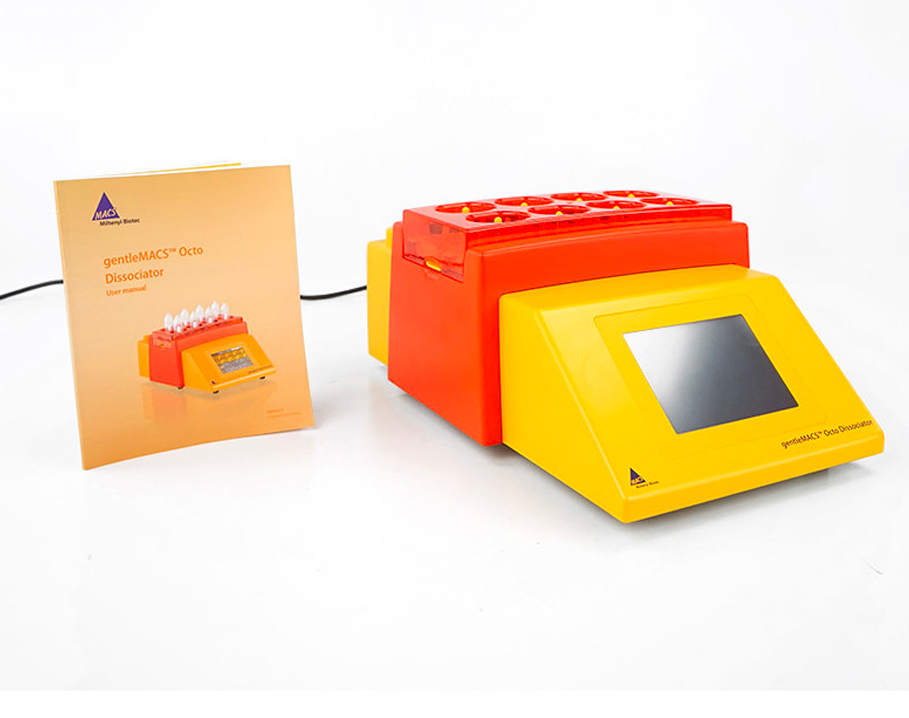
Dissociator Octo GentleMacs (Miltenyi Biotec)
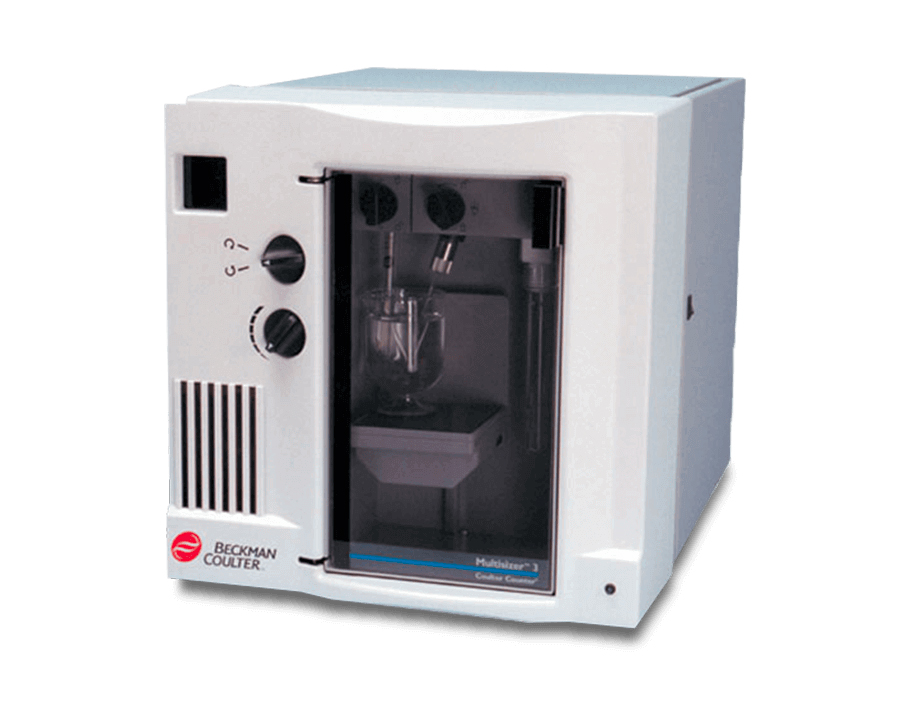
Cell counter Multisizer 3 (Beckman)
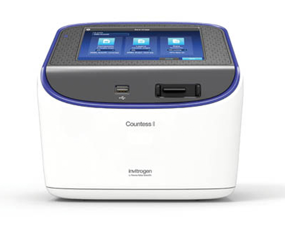
Counter (Thermofisher)
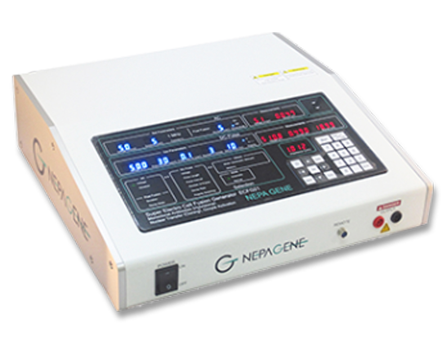
Super electroporator Nepa 21 (Sonidel)
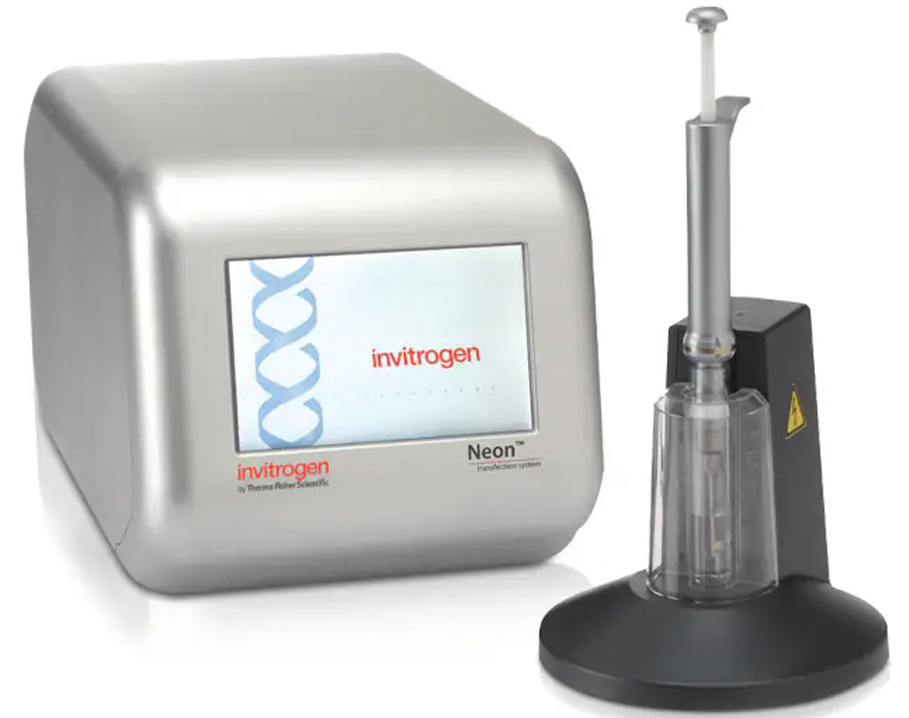
electroporator Neon Trasfection System (Thermofisher)
Gallery


Ilaria Rancati
Head of the Cellular and Preclinical Models Core Facility, IFOM – ETS (AIRC Institute of Molecular Oncology)
Ilaria Rancati leads the Cellular and Preclinical Models Core Facility at IFOM, supporting scientific projects with expertise in cell biology, molecular research, and biorepository operations. She manages biosafety level 1 and 2 cell culture rooms, oversees biorepository, and ensures regulatory compliance.
She designs and delivers training courses on cell culture manipulation and biosafety, fostering expertise within the team. Previously, she specialized in immortalized and primary cell lines and viral vector manipulation. Her technical skills include iPSC generation, CRC organoid culture, neuronal and cardiomyocyte differentiation, and large-scale protein production.
Through her work, Ilaria ensures that researchers have access to high-quality cellular models and training, supporting cutting-edge cancer research.