Tumor Microenvironment and Immunotherapy
Research Description
A tumor doesn’t grow in isolation; it is surrounded by a supportive network of stromal cells, blood vessels and immune cells, forming an ecosystem known as the tumor microenvironment (TME). This environment plays a critical role in shaping cancer development and spreading, ultimately influencing clinical outcomes.
Immunotherapy has transformed the therapeutic landscape of many tumors and can produce exceptional results. Yet, only a minority of patients respond, emphasizing the need to uncover the mechanisms behind treatment success and develop better strategies to activate the immune system.
Importantly, immune cell infiltrates in the TME can either promote or suppress the immune response against the tumor, thereby profoundly influencing response to immunotherapies. With new technologies and tools, researchers can now profile the transcriptomic state and spatial organization of single cells within tumors, revealing the cellular complexity of TMEs with unprecedented resolution.
However, we still lack a comprehensive understanding of how these components function and interact in the evolving TME and how we can evoke effective anti-tumor immune responses to achieve successful immunotherapy outcomes in more patients.
Our lab aims to mechanistically understand and functionally exploit cellular components of the TME to discover new ways to enhance immune responses against tumors. Using patient-derived samples and mouse tumor models, and by employing multiparametric flow cytometry and immunofluorescence, single-cell transcriptomics and in vivo imaging, we investigate: 1) how immune components within the tissue contribute to anti-tumor immune responses, and 2) how suppressive mechanisms develop in tumors and hinder immunotherapy outcomes.
Ongoing Research Projects
1) Tissue-Resident Immune Cells in the TME
Tissues in our bodies harbour different types of resident immune cells which can participate in the cellular composition of the TME. Tissue-resident memory T (Trm) cells normally inhabit peripheral tissues where they play a critical role in protective immunity. They are also strategically positioned to mediate regional tumor surveillance and are important players in the fight against cancer growth.
We are studying how these cells contribute to anti-tumor immunity, how they interact with components of the TME and how they can be harnessed to improve response to immunotherapies.
2) Mechanisms of Immunosuppression in Tumors
Growing tumors adapt to the host immune system by developing resistance mechanisms that suppress anti-tumor immune responses. Regulatory T (Treg) cells are key players of tumor immunosuppression, also by sustaining immunomodulatory myeloid cell states in the TME and dampening immune responses against cancer cells.
We are studying how the suppressive functions of Treg cells are dynamically regulated in tumors and during immunotherapy, and how components of the TME can be leveraged to counteract Treg-mediated responses.

Photogallery
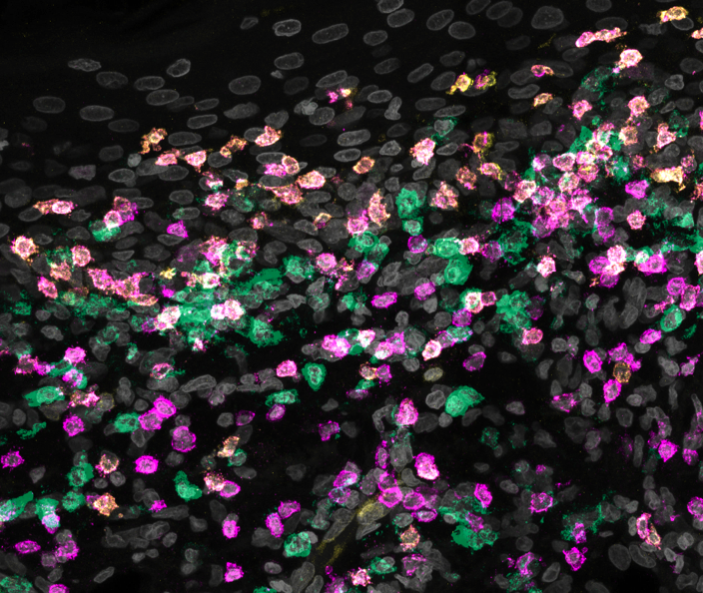
Primary melanoma: confocal microscopy of primary melanoma tissue sample displaying heterogeneous T cells (yellow and pink) infiltrating the tumor and their interactions with antigen presenting cells (green). We are studying how immune components of the tumor microenvironment work together to promote anti-tumor responses.
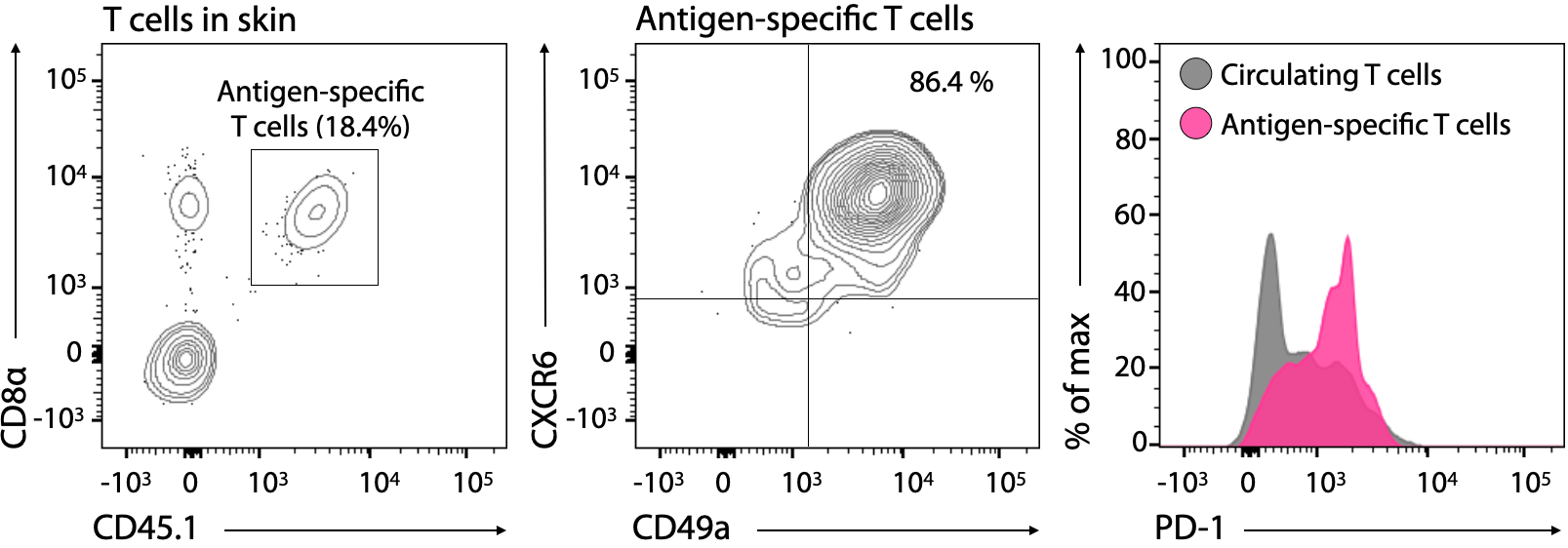
Mouse skin: flow cytometry (FACS) of skin tissue samples derived from mice locally immunized with peptides. T cells specific for antigens of interest are found in the skin as tissue-resident cells and express high levels of PD-1. We are studying how resident T cells in tissues influence tumor progression and immunotherapy outcome.
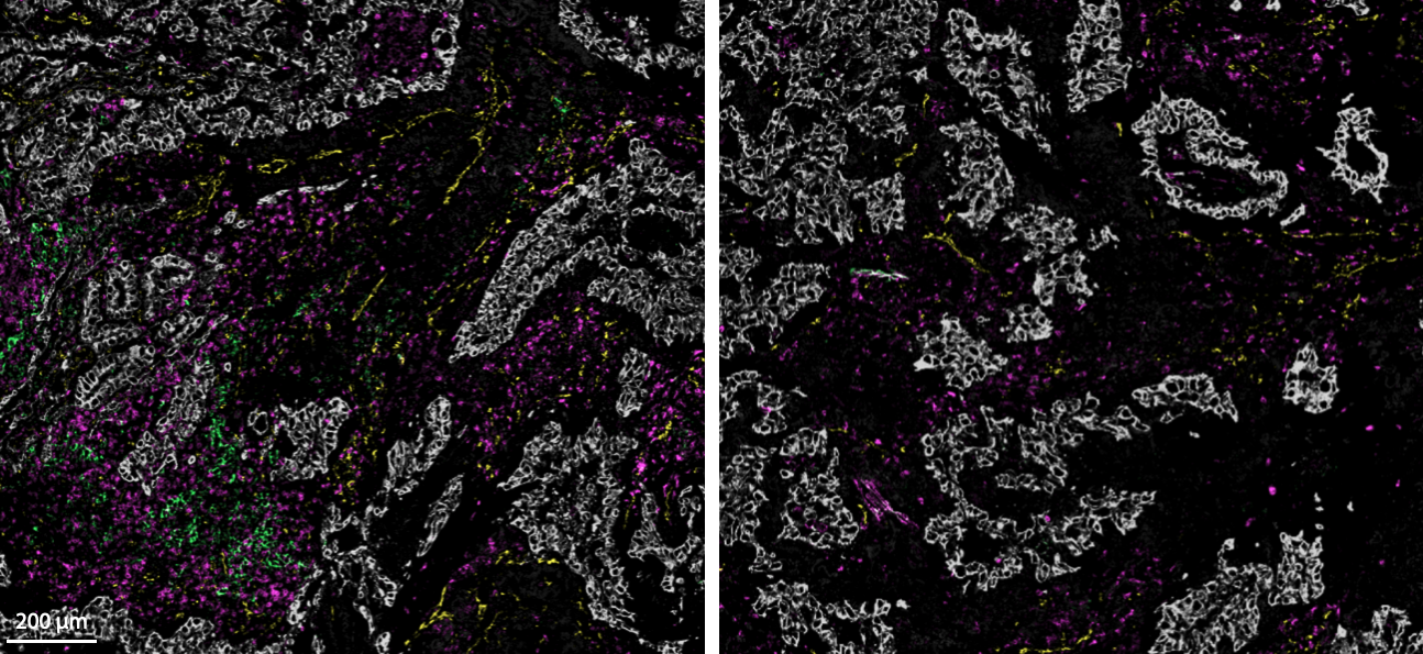
Tumor microenvironment: immunofluorescence of primary non small cell lung cancer (NSCLC) tissue sample displaying different components of the cancer ecosystem including T (pink), myeloid (green), stromal (yellow) and tumor (grey) cells. The two images display differences within the same tissue sample. We are studying how features of the tumor microenvironment are important to drive anti-tumor immunity and successful treatment with immunotherapies.
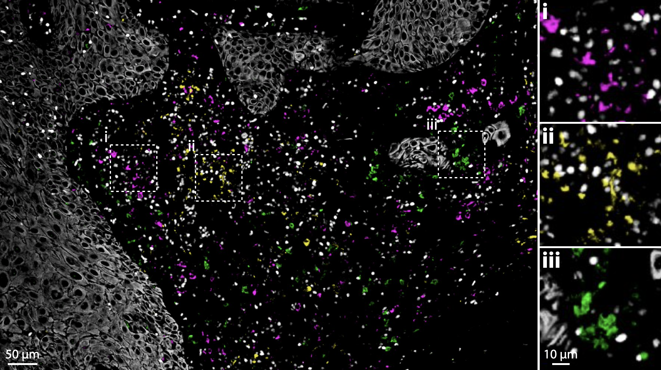
Immunosuppression in tumors: immunofluorescence of primary head and neck squamous cell carcinoma (HNSCC) tissue sample displaying different myeloid cell components including macrophages (pink), activated (yellow) and conventional (green) dendritic cells. All of them can be found near suppressive T cells (white). We are studying how anti-tumor immune responses can be suppressed in the tumor microenvironment during immunotherapy.